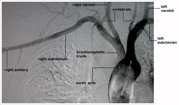
The x-ray dye is injected through a catheter which is located in the aortic arch. Any evaluation of the upper extremity arteries must include an evaluation of the aortic arch and the brachiocephalic trunk.
The aortic arch can be seen, with its three branches: the brachiocephalic trunk, the left common carotid and the left subclavian arteries.
The brachiocephalic trunk divides into the right common carotid and the right subclavian arteries.
The subclavian artery give off several branches, including the vertebral arteries. The subclavian artery becomes the axillary artery at the lateral border of the first rib.